Saturday, November 28, 2015

Morphologic Characteristics of Erythroid Precursors


Pronormoblast (rubriblast):


  • 1% of Nucleated Cells in BM.
  • Size = 20-25 μm.
  • High N/C ratio (8:1)
  • 1-3 faint nucleoli 
  • Lacy chromatin 


Basophilic normoblast (prorubricyte):


  • 1-3% of nucleated cells in BM.
  • Size = 16-18 μm
  • Moderate N/C ratio (6:1)
  • Indistinct nucleoli
  • coarsening chromatin


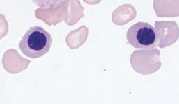
Polychromatophilic normoblast (rubricyte):


  • 13-30% of nucleated cells in BM.
  •  Size = 12 - 15 μm
  • Low N/C ratio (4:1)
  • Chromatin irregular and coarsely clumped
  • Eccentric nucleus


Orthochromic normoblast (metarubricyte):


  • 1-4% of nucleated cells in BM
  • Size = 10 -15 μm
  • Low N/C ratio (1:2)


Reticulocyte (new methylene blue stain):


  • Size = 7-10 μm
  • No N/C ratio because there is no nucleus


Reticulocyte (Wright’s stain):

  • Polychromatophilic (diffusely basophilic)


Mature RBC:


  • Size = 7 -8 μm
  • No nucleus

Thymus


      The thymus is a lymphopoietic organ located in the upper part of the anterior mediastinum. It is a bilobular organ demarcated into an outer cortex and central medulla. The cortex is densely packed with small lymphocytes (thymocytes), cortical epithelial cells, and a few macrophages. The medulla is less cellular and contains more mature thymocytes mixed with medullary epithelial cells, dendritic cells, and macrophages. The primary purpose of the thymus is to serve as a compartment for maturation of T lymphocytes. Precursor T cells leave the bone marrow and enter the thymus through arterioles in the cortex. As they travel through the cortex and the medulla, they interact with epithelial cells and dendritic cells, which provide signals to ensure that T cells can recognize foreign antigen but not self-antigen. They also undergo rapid proliferation. Only about 3% of the cells generated in the thymus exit the medulla as mature T cells. The rest die by apoptosis and are removed by thymic macrophages. The thymus is responsible for supplying the T-dependent areas of lymph nodes, spleen, and other peripheral lymphoid tissue with immunocompetent T lymphocytes.

     The thymus is a well-developed organ at birth and continues to increase in size until puberty. After puberty, however, it begins to atrophy until in old age it becomes barely recognizable. This atrophy could be driven by increased steroid levels beginning in puberty and decreased growth factor levels in adults. The atrophied thymus is still capable of producing new T cells if the peripheral pool becomes depleted as occurs after the lymphoid irradiation that accompanies bone marrow transplantation.


FIGURE: A schematic drawing of the thymus. Hassall’s corpuscles are collections of epithelial cells that may be involved in the development of certain (regulatory) T cell subsets in the thymus. 


Extramedullary Hematopoiesis


     Hematopoiesis in the bone marrow is called medullary hematopoiesis or intramedullary hematopoiesis.

   Blood cell production in hematopoietic tissue other than bone marrow is called extramedullary hematopoiesis.

   In certain hematologic disorders when hyperplasia of the marrow cannot meet the physiologic blood needs of the tissues, extramedullary hematopoiesis can occur in the hematopoietic organs that were active in the fetus, principally the liver and spleen. Organomegaly frequently accompanies significant hematopoietic activity at these sites. This extramedullary hematopoiesis in postnatal life reflects the ability of inert hematopoietic cells to become active, functional cells if the need arises.


Thursday, November 26, 2015

Sites of Hematopoiesis


   Hematopoiesis/Hemopoiesis is the process by whitch blood cells are formed. 

Sites of Hematopoiesis

Yolk sac

     From the 18th day after fertilization, the yolk sac begins hematopoiesis. The cells made here are erythrocytes & few macrophages.

Aorta-gonads-mesonephros (AGM) region

     Located along the developing aorta. This region has the ability to make a wider range of hematopoietic cells including lymphocytes.

    At about the 3rd month of fetal life, the yolk sac & AGM discontinue their role in hematopoiesis.

Liver

    At about the 3rd month of fetal life, the liver becomes the chief site of blood cell production. The liver continues to produce a high proportion of erythroid cells, but myeloid and lymphoid cells begin to appear in greater numbers.

     As fetal development progresses, hematopoiesis also begins to a lesser degree in the spleen, kidney, thymus, and lymph nodes. Erythroid and myeloid cell production as well as early B cell (lymphocyte) development gradually shifts from these sites to bone marrow during late fetal and neonatal life as the hollow cavities within the bones begin to form.

Bone Marrow

     The bone marrow becomes the primary site of hematopoiesis at about the 6th month of gestation and continues as the primary source of blood production after birth and throughout adult life.

    The thymus becomes the major site of T cell (lymphocyte) production during fetal development and continues to be active throughout the neonatal period and childhood until puberty. 

     Lymph nodes and spleen continue as an important site of late B cell differentiation throughout life.

NOTE 

     Liver & spleen may return to hematopoiesis after birth if necessary in a  process  called extramedullary hematopoiesis (production of blood cells outside the bone marrow).


Tuesday, November 24, 2015

Activated Partial Thromboplastin Time (APTT / aPTT / PTT) Test Procedure


Principle

     Platelet-poor plasma is added to an equal volume of partial thromboplastin reagent and warmed to 37°C for an exact incubation time. Pre-warmed (37°C) calcium chloride reagent (0.025M) is added to this mixture to activate the coagulation cascade. The time required for clot formation is recorded. Clot formation may be detected by optical or electromechanical methods using manual, semi-automated, or automated devices.

Reagents and Equipment
  1. Partial thromboplastin reagent: consists of phospholipids and a contact activator
  2. Calcium chloride reagent, 0.025M
  3. Fibrometer system
    a. Fibrometer
    b. Thermal prep block, 37 + 1°C
    c. Automatic pipet
  4. Coagulation cups, non-wettable surface
  5. Fibro-tips
Quality Control

Quality control materials (normal and abnormal) with established control limits should be run. NCCLS recommends controls be tested at the beginning of each testing day, followed by testing during each subsequent shift or with each batch of assays.

Specimen

Whole blood anticoagulated with 3.2% sodium citrate is the specimen of choice. The specimen should be processed to obtain platelet-poor plasma.

Procedure (Fibrometer Testing)
  1. Pre-warm a sufficient quantity of calcium chloride reagent to 37°C for the number of tests to be performed.
  2. Pipet 0.1 mL patient sample or control sample into labeled coagulation cup using the automatic pipet.
  3. To each sample cup, add 0.1 mL of partial thromboplastin reagent using the automatic pipet. Carefully mix the contents of each cup.
  4. Allow sample-partial thromboplastin reagent mixture to pre-warm to 37°C for 1-2 minutes. No longer than five minutes.
  5. Pipet 0.1 mL pre-warmed calcium chloride reagent into coagulation cup containing sample-partial thromboplastin reagent mixture using the automatic pipet (switch in the "on" position). When the calcium chloride reagent is dispensed the timer will automatically start. Alternatively, the timer may be started by touching the timer plate. Reagent should be forcibly added to ensure mixing of reagent and sample.
  6. The timer will stop when clot formation occurs.
  7. Record time taken for clot formation.
  8. Each sample (patient or control) should be run in duplicate.
Results

The average of the duplicate partial thromboplastin times are recorded to the nearest second.

Reference Interval

Each laboratory should establish its own reference interval following a recommended procedure. The reference interval should be re-established with changes in instrumentation, reagent lot number, or at least once a year.

Comments 

1- Partial thromboplastin reagent consists of phospholipids and a contact activator. Kaolin, celite, silica, and ellagic acid are examples of activators available. Care should be taken in choosing the partial thromboplastin reagent, since reagents vary in their sensitivity or insensitivity to lupus anticoagulant.
2- Each laboratory should determine the optimal incubation time for partial thromboplastin reagent and sample based on its assay procedure. The longer the incubation time, the shorter the partial thromboplastin times due to increased contact activation. Excessive heating (>5 minutes) will result in loss of Factor V and Factor VIII.
3- Precision between duplicate measurements is said to be acceptable if the difference between duplicates is 10% or less of the mean of the duplicates.
4- The APTT has been used to monitor heparin therapy. However, newer methodologies such as Factor Xa inhibition assay are replacing this use of the APTT.
5- The APTT is prolonged in:
  • Inherited single factor deficiencies of factors XI, X, IX, VIII, V, II, and I.
  • Disseminated intravascular coagulation (DIC).
  • Presence of circulating inhibitors like lupus-like anticoagulant.
6- If the patient's hematocrit exceeds 55%, NCCLS recommends adjusting the amount of anticoagulant used in the collection tube to prevent over-anticoagulation of the specimen. This correction formula is:

C = (1.85 x 10-3) X (100 - Hct) X V

Where: C = volume of sodium citrate, V = volume of whole blood drawn, Hct = patient's hematocrit.
  

Note
 
Potential sources of error
  • Associated with specimen inappropriate ratio of anticoagulant to blood 
    failure to correct citrate volume if hematocrit >55%
    clotted, hemolyzed, icteric, or lipemic specimen
    delay in processing or testing
    inappropriate storage
  • Associated with reagents incorrect preparation of reagents
    failure to properly store reagents
    use of reagents beyond reconstituted stability time
    use of reagents beyond expiration date
    contaminated reagents
  • Associated with procedure
    incorrect temperature
    incorrect incubation times
    incorrect volumes of sample, reagents, or both
    improperly functioning instrument


Monday, November 23, 2015

Thrombin Time (TT) Test


     TT has an important role as a screening test because it measures the conversion of fibrinogen to fibrin by adding excess thrombin to undiluted plasma. Because the additional clotting factors previously measured in the PT and APTT have no effect on this test, TT is generally useful for evaluating other parameters affecting the formation of fibrin. There can be interference with the conversion of fibrinogen to fibrin for three major reasons: the presence of hypofibrinogenemia or dysfibrinogenemia, the presence of heparin, and the presence of fibrin degradation products (FDP). In rare cases, autoantibodies against thrombin (e.g., induced by topical thrombin application or the use of fibrin sealants) and myeloma proteins can also interfere with fibrin formation and result in an abnormal TT. The TT is useful in corroborating an abnormal FDP result and can verify that the citrated blood sample was drawn through an indwelling heparinized catheter that was not well flushed. An extremely prolonged TT usually indicates a heparin effect. If the sample is contaminated with heparin, it can be absorbed with Hepzyme. The testing can then be repeated, or the specimen can be redrawn.

     The general reference interval for the TT is 10–16 seconds. TT’s sensitivity can be increased by diluting the thrombin reagent to give a control of 16–18 seconds. The TT in preterm and term infants is longer than the adult reference interval even though the fibrinogen level is within the same normal reference interval, which can be explained by the presence of a distinct fetal fibrinogen molecule with altered function. The TT generally becomes normal within a few days after birth.

Thrombin time procedure
  1. Pre-warm 0.1 mL patient or control plasma.
  2. Add 0.2 mL pre-warmed thrombin reagent and start timing device.
  3. Stop timing device upon formation of a clot.
  • Note: Optimal reaction temperature is 37°C. 
  • Reference interval: 10-16 seconds.

NOTE: Conditions associated with prolonged thrombin time


  • Hypofibrinogenemia
  • Dysfibrinogenemia
  • Paraproteins (e.g., cryoglobulin)
  • Presence of heparin
  • Presence of fibrin degradation products
  • Presence of plasmin


Qualitative D-Dimer Test


Principle

     D-dimer is a fibrin fragment that results when plasmin acts on cross-linked fibrin in the presence of factor XIII. D-dimers are produced from an insoluble fibrin clot. Available since the 1990s, this semiquantitative assay provides evidence of normal or abnormal levels of D-dimer. Latex particles are coated with mouse anti–D-dimer monoclonal antibodies. When mixed with plasma containing D-dimers, agglutination occurs. The test plays an important role in detecting and monitoring patients suspected to have thrombotic disorders. Its clinical uses include:

  • Detecting deep vein thrombosis (DVT)
  • Pulmonary embolism (PE)
  • Disseminated intravascular coagulation (DIC)
  • Postoperative complications
  • Septicemia. 

     Quantitative D-dimer procedures are available, using latex-enhanced turbidimetric methods. The qualitative test is widely used in most coagulation laboratories, however, as a screening test for D-dimers.

Reagents and Equipment

1. D-dimer kit containing reagents (stored at 2 to 8°C), good until expiration date on the kit
    a. Test reagent solution containing red blood cell anti–XL-FDP antibody conjugate
    b. Negative control solution containing 0.9% saline solution
    c. Positive control solution containing purified D-dimer fragment
2. Plastic agglutination trays
3. White plastic stirrers
4. Timer
5. 10-μL pipette with disposable tips

Specimen Collection and Storage

1. Collect venous whole blood into a vacuum tube with 3.2% sodium citrate. Dilution of blood to anticoagulant must be 9:1 .
    a. 4.5 mL of blood with 0.5 mL of anticoagulant,  or
    b. 2.7 mL of blood with 0.3 mL of anticoagulant
2. Collection of venous blood into heparin is acceptable.
3. Store specimens at 18 to 24°C. Specimens should be tested within 4 hours of specimen collection. If testing will take place after 4 hours, specimens must be refrigerated at 2 to 8°C; refrigerated specimens are good up to 24 hours.

Quality Control

1. Quality control is performed under several conditions:
    a. Daily
    b. When opening a new kit
    c. When receiving a new shipment
    d. When a new lot number is put into use
2. A whole blood sample that has a negative D-dimer result is used for quality control.
3. Quality control method
    a. Follow directions in the procedure to do the quality control, steps 1 through 5.
    b. Add 1 drop of positive control to the test well, and proceed with steps 6 through 8b.

Procedure

1. Allow reagents to come to room temperature for at least 20 minutes before use.
2. Specimen should be thoroughly mixed; do not allow cells to settle out.
3. For each sample, pipette 10 μL of whole blood into each reaction well; place wells on a plastic agglutination tray; the first is labeled “negative control well” and the second is labeled “test well.”
4. Add 1 drop of the negative control to the negative control well.
5. Add 1 drop of the test reagent to the test well.
6. With a plastic stirrer, mix the contents of each well thoroughly for 3 to 5 seconds, using a different stirrer for each well and spreading the reagent across the entire well surface.
7. To promote agglutination, mix by gentle rocking of the plastic agglutination tray for 2 minutes.
8. At the end of the 2 minutes, observe for the presence of agglutination.
    a. Positive results: Agglutination is present in the test well compared with no agglutination
in the negative control well.
    b. If the negative control well agglutinates, the test is invalid.
    c. If the test result is negative, add 1 drop of positive control to the test well and rock the plastic tray. Agglutination should occur within 15 seconds. If agglutination does not occur with the addition of the positive control, the test is invalid.

Interpretation

1. Positive: Agglutination seen in the test well and no agglutination seen in the negative control well.
2. Negative: No agglutination seen in the test well and the negative control well. This would be confirmed by adding the positive control to the test well and observing agglutination.
3. Invalid
    a. Agglutination occurs in the negative control well.
    b. No agglutination occurs with the positive control.

Results
  • Negative: No agglutination seen in negative agglutination well (less than 0.5 mg/L)
  • Positive: Agglutination seen in undiluted sample (0.5 to 4.0 mg/L)
  • Positive samples can be diluted 1:8 or 1:64 to provide more specific semiquantitative data on the amount of D-dimer present.
Limitations

     The presence of cold agglutinins in patient samples can cause agglutination in patient blood. This may cause agglutination of the negative control, invalidating the test results.
     A quantitative D-dimer test is available, and it is usually performed using a turbidimetric procedure on automated coagulation equipment. The advantage of this method is that it gives an absolute quantity of D-dimer in milligrams per liter, which is an effective tool for determining whether a thrombotic episode has occurred or predicting whether one will occur.


Sunday, November 22, 2015

Prothrombin Time (PT) Manual Test Procedure


Principle

     The addition of pre-warmed (37°C) platelet-poor plasma to thromboplastin-calcium reagent activates the coagulation cascade at Factor VII. The time required for clot formation is recorded. Clot formation may be detected by optical or electromechanical methods using manual, semi-automated, or automated devices.
Reagents and Equipment
  1. Thromboplastin-calcium reagent
  2. Fibrometer system
    a. Fibrometer
    b. Thermal prep block, 37 + 1°C
    c. Automatic pipet
  3. Coagulation cups, non-wettable surface
  4. Fibro-tips
Quality Control

     Quality control materials (normal and abnormal) with established control limits should be run. NCCLS recommends controls be tested at the beginning of each testing day, followed by testing during each subsequent shift or with each batch of assays.

Specimen

     Whole blood anticoagulated with 3.2% sodium citrate is the specimen of choice. The specimen should be processed to obtain platelet-poor plasma.

Procedure (Fibrometer Testing)
  1. Pre-warm a sufficient quantity of thromboplastin-calcium reagent to 37°C for the number of tests to be performed.
  2. Pipet 0.1 mL patient sample or control sample into labeled coagulation cup using the automatic pipet.
  3. Allow sample to pre-warm to 37°C for 1-2 minutes. No longer than five minutes.
  4. Pipet 0.2 mL pre-warmed thromboplastin-calcium reagent into coagulation cup containing patient or control sample using the automatic pipet (switch in the "on" position). When the thromboplastin-calcium reagent is dispensed the timer will automatically start. Alternatively, the timer may be started by touching the timer plate. Reagent should be forcibly added to ensure mixing of reagent and sample.
  5. The timer will stop when clot formation occurs.
  6. Record time taken for clot formation.
  7. Each sample (patient or control) should be run in duplicate.
Results
     The average of the duplicate prothrombin times is recorded to the nearest half -second. To report results in INR, use the following calculation:
INR = (patient's PT/mean PT of the normal range)ISI

Reference Interval

     Each laboratory should establish its own reference interval following a recommended procedure. The reference interval should be re-established with changes in instrumentation, reagent lot number, or at least once a year.

Comments
  1. Precision between duplicate measurements is said to be acceptable if the difference between duplicates is 10% or less of the mean of the duplicate results.
  2. If the patient's hematocrit exceeds 55%, NCCLS recommends adjusting the amount of anticoagulant used in the collection tube to prevent over-anticoagulation of the specimen. This correction formula is:

    C = (1.85 x 10-3) X (100 - Hct) X V

    Where: C = volume of sodium citrate, V = volume of whole blood drawn, Hct = patient's hematocrit.

  3. Significant variation in PT results is observed among different commercial thromboplastin-calcium reagents due to the source of thromboplastin. To normalize these variations, each thromboplastin-calcium reagent has an established international sensitivity index (ISI) that is determined by comparison to the gold standard thromboplastin used by the World Health Organization. The reagent's ISI is used in the calculation of the international normalized ratio (INR). Failure to use the ISI established for a particular lot number of thromboplastin-calcium reagent will result in errors in the INR result. Since the INR is used to monitor patients on oral anticoagulant therapy, an error could lead to misinterpretation of the patient's anticoagulation status.
  4. The prothrombin time is prolonged in individuals with inherited single factor deficiencies of factors X, VII, V, II, I; individuals with liver disease; individuals with vitamin K deficiency; individuals with disseminated intravascular coagulation; and individuals taking oral anticoagulants.

    NOTE:

    Potential sources of error:-

    1. Associated with specimen:- Inappropriate ratio of anticoagulant to blood.
      Failure to correct citrate volume if hematocrit >55% .
      Clotted, hemolyzed, icteric, or lipemic specimen.
      Delay in processing or testing.
      Inappropriate storage.
    2. Associated with reagents:- Incorrect preparation of reagents.
      Failure to properly store reagents.
      Use of reagents beyond reconstituted stability time.
      Use of reagents beyond expiration date.
      Contaminated reagents.
    3. Associated with procedure:- Incorrect temperature.
      Incorrect incubation times.
      Incorrect volumes of sample, reagents, or both.
      Improperly functioning instrument.


Hemacytometer


     Cell counts are performed manually by diluting blood with a diluent, loading a small amount of the diluted sample on a ruled device (hemacytometer), and counting the cells microscopically. The hemacytometer consists of two side-by-side identically ruled glass platforms mounted in a glass holder. Each platform contains a ruled square measuring 3 X 3 mm (9 mm2) and is subdivided according to the improved Neubauer ruling. This ruling subdivides the ruled square into 9 large squares, each measuring 1 X 1 mm (1mm2). All 9 squares are used for leukocyte counts. The large center square is used for platelet and erythrocyte counts. This center square is divided into 25 smaller squares, each with an area of 0.04 mm2. The 5 squares labeled R are used in performing the erythrocyte count whereas the entire center square is used in performing the platelet count.
     On either side of the two ruled glass platforms is a raised ridge. The coverglass is placed on top of the ridge. The distance between the coverglass and the surface of the ruled
area (depth) is exactly 0.1 mm. Thus, the ruled area on each side of the hemacytometer holds a volume of 0.9 mm3 (3 X 3 X 0.1).


     The unopette system is a fast and accurate method for collecting and diluting blood for cell counts. For each laboratory determination performed, the unopette consists of the following elements: the reservoir, the pipet, and the pipet shield. The reservoir contains a premeasured volume of diluting fluid that is specific for the cell count to be performed.


Figure: Unopette system (from left to right): pipet shield, pipet, and reservoir. 


     In summary: 


Figure: Neubauer hemacytometer counting area. The entire counting area is 9 mm2 (3 mm × 3 mm) and is divided into 9 squares. Each square is 1 mm2 (1 mm × 1 mm) in area. Using the 10× objective, 1 square of the counting area (1 mm2) can be viewed. For the manual leukocyte count, all 9 squares are counted. The center square is further divided and the R squares are used for erythrocyte counts, while the entire center square is used for platelet counts.


Anticagulants


Most tests performed in the hematology laboratory involve anticoagulated blood. Once the blood has left the body, a series of reactions occurs causing blood to clot within minutes. To prevent coagulation from occurring, a substance called an anticoagulant is mixed with the blood. Three anticoagulants are used in the hematology laboratory:-
  1. Ethylenediaminetetraacetic acid (EDTA)
  2. Sodium citrate.
  3. Heparin
EDTA:-

EDTA is the most commonly used anticoagulant in the routine hematology laboratory. Sample collection tubes can contain one of three different salt forms of EDTA:
  1. Disodium (Na2EDTA).
  2. Dipotassium (K2EDTA).
  3. Tripotassium (K3EDTA).
The sample collection tube’s stopper is color coded in lavender to indicate the presence of EDTA. EDTA prevents coagulation by chelating calcium, anecessary component of the coagulation cascade. Its removal inhibits the coagulation process. The optimal concentration is 1.5 mg of EDTA per mL blood. Tests using EDTA samples include:
  • Complete blood count (CBC)
  • Hematocrit
  • Peripheral blood smear examination
  • Platelet count,
  • Reticulocyte count,
  • Flow cytometry. 
Ideally, the sample should be used within 6 hours of collection for the majority of these tests. The sample’s stability can be extended to 24 hours with storage at 4°C for certain tests such as the CBC and platelet count. If peripheral blood smears are to be prepared, they should be made within 3 hours of collection. After 3 hours at room temperature, degenerative changes can be observed by examining a Wright-stained blood smear. These changes include:
  • Leukocytes with vacuoles.
  • Irregular cytoplasmic borders.
  • Irregularly shaped nuclei.
  • Platelets increase in size. 
Excess anticoagulant causes erythrocyte shrinkage. Concentrations of more than 2 mg of EDTA per mL blood cause false decreases in the microhematocrit and erythrocyte sedimentation rate (ESR).

Sodium Citrate:-

Sodium citrate is the recommended anticoagulant for coagulation studies. The Clinical and Laboratory Standards Institute (CLSI) recommends the use of 3.2% sodium citrate. The sample collection tube’s stopper is color coded in light blue to indicate the presence of sodium citrate. Sodium citrate prevents coagulation by binding calcium in a soluble complex. The appropriate ratio of anticoagulant: blood is 1:9 for coagulation studies and 1:4 for erythrocyte sedimentation rate (ESR) test.

Heparin:-

Lithium heparin is the CLSI recommended salt of heparin to be used for laboratory testing. The sample collection tube’s stopper is color coded in green to indicate the presence of heparin. Heparin’s interaction with antithrombin prevents coagulation. The interaction leads to the inhibition of thrombin. The recommended concentration for sample collection tubes is 15–30 units heparin/mL blood. Lithium heparin is specifically recommended for the following laboratory tests:
  • Ammonia
  • Carboxyhemoglobin
  • Blood gases
  • Zinc
  • Potassium.
  • Osmotic fragility test.
In hematology, lithium heparin is the appropriate anticoagulant for the osmotic fragility test. The use of heparin for routine hematology procedures is not appropriate. Heparin can affect the platelets and leukocytes, causing them to clump. In addition, heparin causes morphologic distortion of platelets and leukocytes and tends to cause a bluish discoloration of the background of blood films stained with a Romanowsky stain such as Wright stain.

Friday, November 20, 2015

Sickle Cell Screening Procedure


Principle:-

     The sickle screen kit provides a procedure based on hemoglobin solubility. Hemoglobin S is insoluble when combined with a buffer and a reducing agent. This occurs when the blood is mixed with the buffer and sodium hydrosulfite solution. Cells containing hemoglobin S are insoluble and show a turbid cloudy solution. Normal adult hemoglobin A is soluble and produces a transparent solution. The presence of hemoglobin S in either the heterozygous or the homozygous state produces a cloudy solution. Because this is a qualitative screening procedure, all positive results must be followed up with hemoglobin electrophoresis at  alkaline or acid pH or isoelectric focusing.

Reagents and Equipment:-

1. Sickle cell kit.


a. Phosphate buffer/sodium hydrosulfite solution. Prepare by pouring entire contents of sodium hydrosulfite vial into one phosphate buffer bottle. Cap and mix for 1 to 2 minutes. Once reconstituted, reagent is good for 5 days when stored at 2 to 8°C.


b. Unmixed reagents are good until expiration date on package when stored at 2 to 8°C.


2. 12-mm 75-mm test tubes.


3. Test tube caps or parafilm.


4. 50-μL pipette and tips.


5. Reading rack.


6. 5-mL pipette (to pipette the buffer).

Specimen Collection and Storage:-

1. Whole blood obtained in EDTA, heparin, or sodium citrate.


2. Specimens can be refrigerated at 2° to 8°C for 2 weeks before testing.

Quality Control:-

Commercially prepared negative and positive controls are run along with the patient’s blood. Control results must be correct to report patient results.

Procedure:-

1. Pipette 4 mL of the phosphate buffer/sodium hydrosulfite solution to each test tube (one for each test and each control).


2. Add 50 μL of well-mixed whole blood or control to each labeled tube.


3. Cover each tube with a cap or parafilm, and invert to mix three or four times.


4. Place each tube in the reading rack at room temperature, and let the tubes incubate for 10 to 20 minutes.

Interpretation of Results and Result Reporting:-


Positive: If hemoglobin S or any other sickling hemoglobin (hemoglobin C Harlem) is present, the solution will be turbid and the lines on the reading rack will be invisible.
Negative: If no sickling hemoglobin is present, the lines on the reading rack will be visible through the solution (figure below).


 Limitations:-

1. Severe anemias can cause false-negative results. The  sample volume should be doubled (100 μL) if the hemoglobin is less than 8 g/dL.

2. False-negative results can occur with infants younger than 6 months because hemoglobin F is insoluble in the test solution. Therefore, testing should not be done on infants.


3. Patients with multiple myeloma, cryoglobulinemia, and other dysglobulinemias may show false-positive results because the high protein level may affect the test.


4. Some rare hemoglobin variants such as hemoglobin C Harlem or C Georgetown may give false-positive results. These are sickling hemoglobins but do not contain hemoglobin S.


5. Patients who have been recently transfused may give false-positive or false-negative results.


6. Patients who have sickle trait give positive results. These results should be confirmed with hemoglobin electrophoresis.


7. Positive results and questionable results should be confirmed with hemoglobin electrophoresis.

Thursday, November 19, 2015

Functions of the Spleen

Hematopoietic function:-

  • Can produce white blood cells, red blood cells, and platelets if necessary.

Reservoir function:-

  • One-third of platelets and granulocytes are stored in the spleen.

Filtration functions:-

  • Aging red blood cells are destroyed.
  • Spleen removes inclusion from red blood cells.
  • If red blood cell membrane is less deformable or antibodycoated, spleen presents a hostile environment leading to production of spherocytes.

Immunologic function:-

  • Opsonizing antibodies are produced, trapping and processing antigens from encapsulated organs.


Saturday, November 7, 2015

Lineage-specific Cytokine Regulation


LINEAGE-SPECIFIC CYTOKINE REGULATION

Erythropoiesis:

In the erythroid lineage, progenitor cells give rise to two distinct types of erythroid colonies in culture. A primitive progenitor cell, the BFU-E, is relatively insensitive to EPO and forms large colonies after 14 days in the form of bursts. Production of BFU-E colonies was originally described as being supported by burst-promoting activity, or BPA, now known to be IL-3 or GM-CSF. CFU-E colonies grow to maximal size in 7 to 8 days and depend primarily on EPO. The CFU-E are the descendants of BFU-E and subsequently give rise to the first recognizable erythrocyte precursor, the pronormoblast. Other cytokines reported to influence production of red cells include IL-9, IL-11, and SCF. However, EPO is the pivotal humoral factor that functions to prevent apoptosis and induce proliferation/differentiation of the most committed erythroid progenitor cells and their progeny.

Granulopoiesis and Monopoiesis:

Granulocytes and monocytes are derived from a common bipotential progenitor cell, the CFU-GM, derived from CFUGEMM. Specific GFs for granulocytes and monocytes, acting synergistically with GM-CSF and/or IL-3, support the differentiation pathway of each lineage. M-CSF supports monocyte differentiation while G-CSF induces neutrophilic granulocyte differentiation. Eosinophils and basophils also are derived from the CFU-GEMM under the influence of growth factors IL-5 and IL-3/IL-4, respectively.

Megakaryocytopoiesis/Thrombopoiesis:

Platelets are derived from megakaryocytes, which are progeny of the CFU-EMk. CFU-Mk are induced to proliferate and differentiate into megakaryocytes by several cytokines. However, the cytokines that induce the greatest increase in platelet production are IL-11 and TPO.

Lymphopoiesis:

The growth and development of lymphoid cells from the common lymphoid progenitor cell occurs in multiple anatomic locations including the bone marrow, thymus, lymph nodes, and spleen. Multiple GFs play a role in T and B lymphocyte growth and development, most of which act synergistically.