Multiple myeloma (myelomatosis) is a neoplastic disease characterized by plasma cell accumulation in the bone marrow, the presence of monoclonal protein in the serum and/or urine and, in symptomatic patients, related tissue damage.
Friday, December 4, 2015
Reticulocyte
Reticulocyte ( Polychromatic Erythrocyte / Diffusely Basophilic Erythrocyte )
- Size = 7-10 μm
- No nucleus (so there is no N/C ratio)
- No nucleoli
- No chromatin
- Cytoplasm: Color is slightly more blue/purple than the mature erythrocyte because there are RNA remnants.
- Bone marrow: 1 %
- Peripheral blood: 0.5 - 2.0 %
NOTE: When stained with supravital stain (e.g., new methylene blue), polychromatic erythrocytes appear as reticulocytes (contain precipitated ribosomal material)
Thursday, December 3, 2015
Orthochromic Normoblast
Orthochromic Normoblast ( Orthochromic Erythroblast / Metarubricyte )
- 1 - 4% of nucleated cells in BM
- Peripheral blood: 0 %
- Size = 10 -15 μm
- Low N/C ratio (1:2)
- Round nucleus
- No nucleoli
- Fully condensed chromatin
- Cytoplasm: more pink or salmon than blue
NOTE: The gray-blue color of the cytoplasm is becoming salmon as more hemoglobin is produced.
Wednesday, December 2, 2015
Polychromatophilic Normoblast
Polychromatophilic Normoblast ( Polychromatic Normoblast / Polychromatic Erythroblast / Rubricyte )
- 13-30% of nucleated cells in BM.
- Peripheral Blood : 0 %
- Size = 12 - 15 μm
- Low N/C ratio (4:1)
- Eccentric nucleus
- No nucleoli
- Chromatin irregular and coarsely clumped
- Cytoplasm: Gray-blue as a result of hemoglobinization
NOTE: The blue color of the cytoplasm is becoming gray-blue as hemoglobin is produced.
Tuesday, December 1, 2015
Basophilic Normoblast
Basophilic Normoblast (Basophilic Erythroblast / Prorubricyte)
- 1-3% of nucleated cells in BM.
- Peripheral blood : 0 %
- Size = 16-18 μm
- Round to slightly oval nucleus
- Moderate N/C ratio (6:1)
- Dark blue cytoplasm
- Indistinct nucleoli ( 0 - 1 nucleoli )
- Coarsening ( slightly condensed ) chromatin
Pronormoblast
Pronormoblast (also known as Proerythroblast / Rubriblast) is the first microscopically recognizable cell in erythrocyte lineage.
- 1% of Nucleated Cells in BM.
- Peripheral Blood: 0%
- 1% of Nucleated Cells in BM.
- Size: 20-25 μm
- Nucleus: Round to slightly oval
- High N/C ratio (8:1)
- 1-3 faint nucleoli
- Fine chromatin
- Cytoplasm: Dark blue; may have prominent Golgi
Glycosylated Hemoglobin
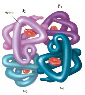
HbA1C on chromatography is a minor component of normal adult hemoglobin (HbA) that has been modified posttranslationally (HbA3 on starch block electrophoresis). A component usually has been added to the N terminus of the β-chain. The most important subgroup of HbA1 is HbA1C , which has glucose irreversibly attached. This hemoglobin is referred to as glycosylated hemoglobin. HbA1C is produced throughout the erythrocyte’s life, its synthesis dependent on the concentration of blood glucose. Older erythrocytes typically contain more HbA1C than younger erythrocytes having been exposed to plasma glucose for a longer period of time. However, if young cells are exposed to extremely high concentrations of glucose ( >400 mg/dL) for several hours, the concentration of HbA1C increases with both concentration and time of exposure.
Measurement of is routinely used as an indicator of control of blood glucose levels in diabetics because it is proportional to the average blood glucose level over the previous two to three months.
Measurement of is routinely used as an indicator of control of blood glucose levels in diabetics because it is proportional to the average blood glucose level over the previous two to three months.
Average levels of HbA1C are 7.5% in diabetics and 3.5% in normal individuals.


